
Scientific Volume Imaging
Experts in microscopic imaging
Scientific Volume Imaging
develops high quality restoration
(deconvolution) and visualization software for microscopists all over
the world.
One single Huygens software license for deconvolution and visualisation
software can be run simultaneously as often as needed from all computers
in a network via a simple X-server tool
(http://www.svi.nl/solutions/display.php).
Running on five platforms, 64
bit since 1999 and with good memory management Huygens can handle the
largest datasets with consummate ease for Widefield, Brightfield,
Confocal, Multi-photon, Spinning-disc and 4Pi microscopes. With its
native 2D,3D and 4D file reading in ICS, ICS2, all types of Tiffs, Leica
Tiffs, Zeiss LSM5, Metamorph STK, MRC, Olympus FluoView, BioVision and
DeltaVision Huygens gives a smooth and complete follow up on the
acquisition software delivered with your microscope.
Some key features:
- Runs on Windows, Linux, Mac, Irix SGI and AIX IBM.
- Three basic packages (Essential, Professional and Scripting) ranging
from a easy Wizard approach until fully automated deconvolution of
multiple imagestacks can all share the same options.
- Supports multiprocessing, 64 bit.
- Automatic Z-drift, bleaching correction and correction for spherical
abberation.
- Automatic measurement of various depth dependent PSF's based either on
beads or the parameter information.
- Many visualisation tools right under your mouse button: Surface &
Volume rendering, Twin slicer, MIP renderer.
- Analysis tools for all kinds of measurement and Colocalisation
- Free support and updates for a full year.
SVI's dedicated team of programmers stays in close contact with the
international scientific microscopic community to continuously improve
and expand Huygens.
Therefore SVI has set up the SVI-Wiki. This is a rapidly expanding public knowledge resource on 3D microscopy and deconvolution. Based on the WikiWikiWeb principle, it is open to contributions from every visitor. In addition it serves as a support medium for SVI customers and relations to discuss different aspects of SVI's Huygens Software.The SVI-wiki can be found in http://support.svi.nl/wiki/
Deconvolution & Visualization Software

|
Huygens® Professional
Discover the truth behind your image!
With Huygens® Professional you can restore images without being a
specialist on image restoration and optical theory. For instance, intelligent
tools make it possible for you to obtain the instrumental Point Spread
Function of your microscope from an easily recorded latex bead image. This
enables you to make sure that your microscope is indeed functioning optimally
and ensures optimal restoration results.
|
|
Huygens® Essential
Deconvolution a few mouse clicks away!
Huygens® Essential is an image processing software package tailored for
deconvolution of microscopic images. Its wizard driven user interface guides
you through the process of deconvolving microscopic images. Huygens Essential
is also equipped with a special tool that enables you to go through the volume
image slice by slice and a tool to extract an PSF from images. Essential can
also be extended with the two visualization options Surface Renderer and
Colocalization Tool.
|

|
Huygens® Scripting
Process your images with automated scripts!
Once you know how to deal with a particular kind of dataset and are sure of
the parameters, you can run a couple or more of the datasets through
Huygens® Scripting. This is called batch processing. The Huygens Scripting
enables you to run scripts written in Tcl, using the extensive set of Huygens
image processing commands. With Huygens Scripting you may construct programs
that launch multiple parallel image processing jobs to make full usage of a
multiple cpu system.
|
Free Visualization Software

|
FreeSFP
FREE Volume Rendering with one push on a button!
FreeSFP is a free of charge volume renderer that enables you to explore 3D
digital images from a selectable viewpoint. It is especially suitable for
single or multi-channel images from microscopes or MRI. The computational work
is done by the Simulated Fluorescence Process (SFP) algorithm that takes the
volume image as a distribution of fluorescent material, simulating what
happens if the material is excited and how the subsequently emitted light
travels to the observer.
|
Usable images
|
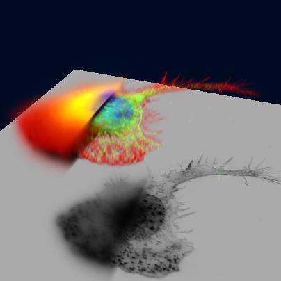
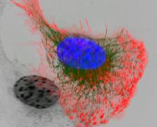
Macrophage fluorescently stained for tubulin (yellow/green), actin (red) and
the nucleus (DAPI, blue) recorded with a widefield microscope. Visualized by
FluVR's spectral fluorescence volume renderer.
Left part: original data.
Right part: as deconvolved with Huygens Professional.
Recorded by Dr. James Evans.
|
|
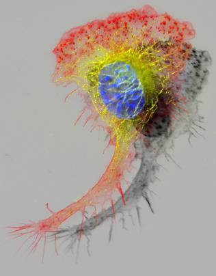
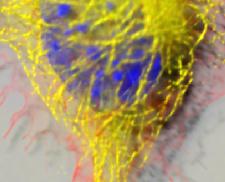
Macrophage fluorescently stained for tubulin (yellow/green), actin (red) and
the nucleus (DAPI, blue) recorded with a widefield microscope as deconvolved
with Huygens Professional and visualized by FluVR's spectral fluorescence
volume renderer.
Recorded by Dr. James Evans.
|
|
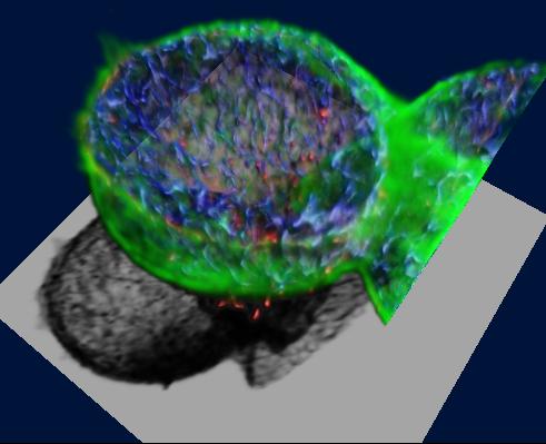
This specimen is an isolated Rat Hepatocyte couplet as deconvolved with Huygens Professional and visualized with FluVR's SFP Rendering engine.
Recorded by Lab of Prof. Lukas Landmann.
|

|
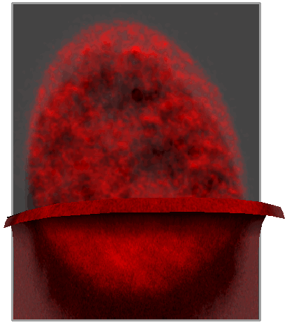
A metaphase human cell stained for DNA (red), centromeres (blue) and the
anaphase promoting complex/cyclosome (green). Visualized by FluVR's spectral
fluorescence volume renderer.
Upper part: original data.
Lower part: deconvolved with Huygens Professional.
Recorded by Dr. Claire Acquaviva, Dr. Pines Lab.
|

|
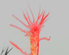
Tube-like intranuclear lamin structure traversing the nucleus of a living cell as visualized with green fluorescent protein (GFP). Original data deconvolved with the Huygens System and volume rendered with the GSFP renderer of FluVR.
Data Courtesy: Dr. J.Broers
|
|
Nucleus of a human epithelium cell stained with an antibody against splicing
factor.
Top part: image as restored by Huygens Professional.
Bottom part: original image.
Both top and bottom images were visualized using the Generalized
Simulated Fluorescence Process (GSFP) volume rendering method.
Recorded by Dr. Marjolein A. Grande
|
|
|
Close ups of Macrophage as deconvolved by Huygens Professional, recorded by
Dr. James Evans
|
Product flyers